SUN Colonoscopy Video Database
Update
- 2022 10/08 : Added supplementary data: Accuracy of the latest approved model (July 2022).
- 2022 07/11 : Revised notations of polyp location in Table 2.
- 2020 10/07 : Released SUN Colonoscopy Video Database.
Abstract
SUN (Showa University and Nagoya University) Colonoscopy Video Database is the colonoscopy-video database for the evaluation of an automated colorectal-polyp detection.
The database comprises of still images of videos, which are collected at the Showa University Northern Yokohama Hospital. Mori Laboratory, Graduate School of Informatics, Nagoya University developed this database.
Every frame in the database was annotated by the expert endoscopists at Showa University.
Summary of database
The SUN database includes 49,136 polyp frames taken from different 100 polyps, which were fully annotated with bounding boxes.
Non-polyp scenes of 109,554 frames are also included in this database. The characteristics of the database are summarized in Tables 1-3.
In polyp-exsiting frames, each polyp is annotated with a bounding box as shown in Figure 1.
The file formats of images, and bounding boxes are jpeg and a text file, respectively.
In the text file, each row represents a bounding box of a polyp, that is,
"Fielename min_Xcoordinate,min_Ycoordinate,max_Xcorrdinate,max_Ycoordinate,class_id",
where class_id of 0 and 1 represent polyp and non-polyp frames, respectively.
Here are an examples:
polyp1_00001.jpg 50,100,150,200,0
polyp1_00002.jpg 120,300,250,600,0
...
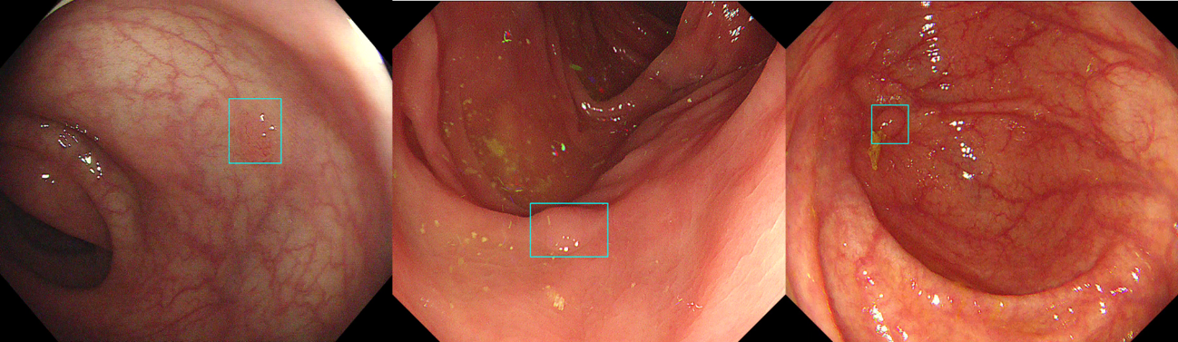 Figure 1: Examples of annotated images. The information of the bounding box is provided by a text file besides image files.
| Patients registered as SUN database (n = 99) | |||
|---|---|---|---|
| Sex (Male / Female) | 71 / 28 | ||
| Median Age (IQR) | 69 (58 – 74) | ||
| Lesions registered as SUN database (n = 100) | |||
| Median Size (IQR) mm | 5 (3 – 7) | ||
| Number of diminutive polyp (≤5mm) | 60 | ||
| Morphology (protruded / flat) | 66 / 34 | ||
| Location (Right / Left / Rectum) | 47 / 44 / 8 | ||
| Pathological diagnosis | |||
| Hyperplastic polyp | 7 | ||
| Sessile serrated lesion | 4 | ||
| Low grade adenoma | 82 | ||
| Traditional serrated adenoma | 2 | ||
| High grade adenoma | 4 | ||
| Invasive carcinoma | 1 | ||
| ID | Number of frames | Shape | Size | Location | Pathological diagnosis |
|---|---|---|---|---|---|
| 1 | 527 | Is | 6mm | Cecum | Low-grade adenoma |
| 2 | 1,313 | Is | 18mm | Rectum | High-grade adenoma |
| 3 | 292 | IIa | 3mm | Ascending colon | Low-grade adenoma |
| 4 | 80 | Is | 4mm | Sigmoid colon | Low-grade adenoma |
| 5 | 930 | IIa | 3mm | Transverse colon | Low-grade adenoma |
| 6 | 491 | IIa | 3mm | Sigmoid colon | Low-grade adenoma |
| 7 | 315 | IIa | 6mm | Descending colon | Low-grade adenoma |
| 8 | 256 | Isp | 12mm | Sigmoid colon | Low-grade adenoma |
| 9 | 136 | Is | 4mm | Sigmoid colon | Low-grade adenoma |
| 10 | 436 | IIa | 3mm | Transverse colon | Low-grade adenoma |
| 11 | 113 | IIa | 5mm | Descending colon | Low-grade adenoma |
| 12 | 538 | Is | 5mm | Rectum | Low-grade adenoma |
| 13 | 479 | Is | 5mm | Transverse colon | Low-grade adenoma |
| 14 | 1,183 | IIa | 3mm | Sigmoid colon | Low-grade adenoma |
| 15 | 487 | Is | 5mm | Transverse colon | Low-grade adenoma |
| 16 | 199 | Is | 4mm | Transverse colon | Low-grade adenoma |
| 17 | 304 | Is | 4mm | Sigmoid colon | Low-grade adenoma |
| 18 | 243 | Is | 2mm | Sigmoid colon | Hyperplastic polyp |
| 19 | 96 | IIa | 3mm | Transverse colon | Low-grade adenoma |
| 20 | 3159 | IIa | 3mm | Ascending colon | Low-grade adenoma |
| 21 | 100 | IIa | 3mm | Sigmoid colon | Low-grade adenoma |
| 22 | 314 | IIa | 2mm | Ascending colon | Low-grade adenoma |
| 23 | 182 | Ip | 12mm | Ascending colon | Low-grade adenoma |
| 24 | 973 | Ip | 15mm- | Sigmoid colon | Low-grade adenoma |
| 25 | 338 | Is | 7mm | Sigmoid colon | Low-grade adenoma |
| 26 | 370 | Is | 5mm | Descending colon | Low-grade adenoma |
| 27 | 249 | Is | 5mm | Ascending colon | Hyperplastic polyp |
| 28 | 195 | Is | 2mm | Transverse colon | Low-grade adenoma |
| 29 | 377 | Isp | 13mm | Sigmoid colon | Low-grade adenoma |
| 30 | 224 | IIa | 4mm | Sigmoid colon | Low-grade adenoma |
| 31 | 183 | Ip | 12mm | Descending colon | Low-grade adenoma |
| 32 | 981 | Ip | 15mm- | Ascending colon | Traditional serrated adenoma |
| 33 | 594 | Is | 5mm | Sigmoid colon | Low-grade adenoma |
| 34 | 245 | Is | 3mm | Ascending colon | Low-grade adenoma |
| 35 | 1,212 | Ip | 15mm- | Sigmoid colon | High-grade adenoma |
| 36 | 815 | IIa | 7mm | Sigmoid colon | Low-grade adenoma |
| 37 | 448 | Is | 7mm | Transverse colon | Low-grade adenoma |
| 38 | 509 | Is | 5mm | Ascending colon | Low-grade adenoma |
| 39 | 713 | IIa | 13mm | Ascending colon | Low-grade adenoma |
| 40 | 159 | IIa | 5mm | Transverse colon | Low-grade adenoma |
| 41 | 108 | IIa | 3mm | Rectum | Low-grade adenoma |
| 42 | 268 | Is | 7mm | Transverse colon | Low-grade adenoma |
| 43 | 260 | Isp | 10mm | Ascending colon | Low-grade adenoma |
| 44 | 745 | IIa | 5mm | Sigmoid colon | Low-grade adenoma |
| 45 | 383 | Is | 3mm | Ascending colon | Low-grade adenoma |
| 46 | 170 | IIa | 2mm | Transverse colon | Hyperplastic polyp |
| 47 | 705 | Is | 5mm | Transverse colon | Low-grade adenoma |
| 48 | 176 | Is | 3mm | Transverse colon | Low-grade adenoma |
| 49 | 181 | IIa | 3mm | Transverse colon | Low-grade adenoma |
| 50 | 740 | Ip | 10mm | Sigmoid colon | Low-grade adenoma |
| 51 | 1,737 | IIa(LST-NG) | 15mm- | Cecum | Low-grade adenoma |
| 52 | 207 | IIa | 6mm | Sigmoid colon | Low-grade adenoma |
| 53 | 245 | Is | 4mm | Rectum | Hyperplastic polyp |
| 54 | 345 | Is | 4mm | Sigmoid colon | Low-grade adenoma |
| 55 | 700 | Is | 3mm | Ascending colon | Low-grade adenoma |
| 56 | 248 | Is | 4mm | Sigmoid colon | Hyperplastic polyp |
| 57 | 326 | Is | 5mm | Transverse colon | Low-grade adenoma |
| 58 | 267 | IIa | 6mm | Transverse colon | Sessile serrated lesion |
| 59 | 646 | Isp | 8mm | Sigmoid colon | Traditional serrated adenoma |
| 60 | 146 | IIa | 8mm | Transverse colon | Low-grade adenoma |
| 61 | 679 | Isp | 6mm | Ascending colon | Low-grade adenoma |
| 62 | 351 | Is | 7mm | Ascending colon | Low-grade adenoma |
| 63 | 632 | Is | 7mm | Rectum | Invasive cancer (T1b) |
| 64 | 81 | IIa | 3mm | Sigmoid colon | Low-grade adenoma |
| 65 | 222 | IIa | 3mm | Cecum | Low-grade adenoma |
| 66 | 1,685 | Is | 6mm | Sigmoid colon | Low-grade adenoma |
| 67 | 191 | IIa | 5mm | Transverse colon | Low-grade adenoma |
| 68 | 1319 | Is | 15mm- | Rectum | High-grade adenoma |
| 69 | 130 | IIa | 3mm | Descending colon | Low-grade adenoma |
| 70 | 264 | Ip | 15mm- | Sigmoid colon | Low-grade adenoma |
| 71 | 1,021 | Is | 4mm | Ascending colon | Low-grade adenoma |
| 72 | 774 | Is | 5mm | Ascending colon | Low-grade adenoma |
| 73 | 1,285 | Is | 3mm | Cecum | Low-grade adenoma |
| 74 | 276 | Isp | 5mm | Sigmoid colon | Low-grade adenoma |
| 75 | 343 | Is | 3mm | Transverse colon | Low-grade adenoma |
| 76 | 343 | Is | 3mm | Cecum | Low-grade adenoma |
| 77 | 215 | Is | 4mm | Ascending colon | Low-grade adenoma |
| 78 | 267 | Isp | 12mm | Sigmoid colon | High-grade adenoma |
| 79 | 76 | Is | 4mm | Descending colon | Low-grade adenoma |
| 80 | 1,192 | Is | 10mm | Sigmoid colon | Low-grade adenoma |
| 81 | 427 | Is | 6mm | Sigmoid colon | Low-grade adenoma |
| 82 | 111 | IIa | 3mm | Sigmoid colon | Sessile serrated lesion |
| 83 | 795 | Isp | 13mm | Rectum | Low-grade adenoma |
| 84 | 218 | Is | 5mm | Descending colon | Low-grade adenoma |
| 85 | 1,393 | IIa | 8mm | Ascending colon | Low-grade adenoma |
| 86 | 257 | IIa | 4mm | Sigmoid colon | Low-grade adenoma |
| 87 | 454 | Is | 3mm | Cecum | Low-grade adenoma |
| 88 | 249 | Is | 4mm | Ascending colon | Low-grade adenoma |
| 89 | 149 | Ip | 5mm | Descending colon | Low-grade adenoma |
| 90 | 479 | Is | 10mm | Ascending colon | Sessile serrated lesion |
| 91 | 1,061 | IIa | 13mm | Ascending colon | Low-grade adenoma |
| 92 | 391 | Is | 7mm | Descending colon | Low-grade adenoma |
| 93 | 452 | Is | 7mm | Descending colon | Low-grade adenoma |
| 94 | 136 | Is | 6mm | Sigmoid colon | Low-grade adenoma |
| 95 | 606 | Isp | 8mm | Sigmoid colon | Low-grade adenoma |
| 96 | 301 | Is | 5mm | Sigmoid colon | Hyperplastic polyp |
| 97 | 431 | IIa | 15mm- | Cecum | Sessile serrated lesion |
| 98 | 170 | IIa | 4mm | Transverse colon | Low-grade adenoma |
| 99 | 161 | Is | 5mm | Sigmoid colon | Low-grade adenoma |
| 100 | 188 | IIa | 3mm | Rectum | Hyperplastic polyp |
| ID | Number of frames | Lenth of each video (seconds) |
|---|---|---|
| 1 | 9,961 | 332.0 |
| 2 | 10,073 | 335.8 |
| 3 | 7,152 | 238.4 |
| 4 | 14,635 | 487.8 |
| 5 | 7,916 | 263.9 |
| 6 | 17,046 | 511.4 |
| 7 | 5,636 | 169.1 |
| 8 | 2,568 | 85.6 |
| 9 | 9,522 | 317.4 |
| 10 | 7,086 | 236.2 |
| 11 | 4,832 | 161.1 |
| 12 | 6,799 | 226.6 |
| 13 | 6,328 | 210.9 |
Supplementary data
Accuracy of the latest approved model (July 2022)
After the publication (Misawa M et al. Gastrointest Endosc 2021;93(4):960-967e3), we updated the EndoBRAIN-EYE (CADe). Therefore, we conducted re-analysis using the latest model to show current performance. The following tables show the latest performances for SUN Colonoscopy Video Database.
| Latest model* | Reported model† | |||
|---|---|---|---|---|
| Percent(95% confidence interval) | n/N | Percent | n/N | |
| Sensitivity (per-lesion) | 98.0 (93.0-99.8) | 98/100 | 98.0 (93.0-99.8) | 98/100 |
| Sensitivity (per-frame) | 91.5 (91.2-91.7) | 44,092/48212 ‡,§ | 90.5 (90.2-90.7) | 44,472/49140 ‡ |
| Specificity | 98.2 (98.1-98.2) | 90,068/91,764 ‡,§ | 93.7 (93.5-93.8) | 88,075/94,039 ‡ |
| *The latest model was regulatory approved in July 2022. | ||||
| †Refer to the Misawa M et al. Gastrointest Endosc 2021;93(4):960-967e3. | ||||
| ‡The numbers of frames were inconsistent since the database was released with the removal of inappropriate frames after the paper publication. | ||||
| §The frames which were identified as inappropriate frames by the CADe system were excluded from the analysis. | ||||
| Total number of frames | True positive* | False negative | False positive† | Unanalyzable frames‡ |
|---|---|---|---|---|
| 48,344 | 44,902 | 3,964 | 156 | 132 |
| *Number of the frames in which the IoU between the predicted bounding box and ground truth more than or equals to 0.3. | ||||
| †Number of the frames to which the trained model outputs bounding box but the IoU less than 0.3. | ||||
| ‡Number of the frames which were identified as inappropriate frame by the CADe system. | ||||
| Total number of frames | False positive | True negative | Unanalyzable framese* |
|---|---|---|---|
| 109,516 | 1,696 | 90,068 | 17,752 |
| *Number of the frames which were identified as inappropriate frame by the CADe system. | |||
| Number of polyp videos | True positive | False negative |
|---|---|---|
| 100 | 98 | 2 |
Terms of use
Copyright
All intellectual property rights, including copyrights, relating to the information contained in this database are held by Showa University Northern Yokohama Hospital and Mori Lab., Graduate School of Informatics, Nagoya University.
Intended use
This database is available for only non-commercial use in research or educational purpose. As long as you use the database for these purposes, you can edit or process images and annotations in this database. Without permission from Mori Lab., commercial use of this dataset is prohibited even after copying, editing, processing or any operations of this database. Please contact us for commercial use or if you are uncertain about the decision.
Citation
If you clearly indicate the following information, you are allowed to refer to or cite the part of this database. By downloading and using the SUN database you agree to cite this database in any publication based on research in which this database has been used.
- Development of a computer-aided detection system for colonoscopy and a publicly accessible large colonoscopy video database (with video). Masashi Misawa, Shin-ei Kudo, Yuichi Mori, Kinichi Hotta, Kazuo Ohtsuka, Takahisa Matsuda, Shoichi Saito, Toyoki Kudo, Toshiyuki Baba, Fumio Ishida, Hayato Itoh, Masahiro Oda, Kensaku Mori, Gastrointestinal Endoscopy, Vol. 93, Issue 4, pp. 960-967.e3, 2021. DOI: 10.1016/j.gie.2020.07.060
- SUN Colonoscopy Video Database. Hayato Itoh, Masashi Misawa, Yuichi Mori, Masahiro Oda, Shin-Ei Kudo, Kensaku Mori, 2020, http://amed8k.sundatabase.org/
Distribution
It is prohibited to sell, transfer, lend, lease, resell, distribute, etc., as it is, or copy, edit, or process this database, in whole or in part.
Personal information
The use of personal information sent to issue the download path of this database is limited to the notifications about this database and usage statistics of this database. Management of information follows the Nagoya University Privacy Policy. Note that this website uses Google Analytics with Cookie for analysis of its accesses. If you want to deny this analysis, please disable Cookie in your webbrowser.
Immunity
Showa University, Nagoya University, and Mori Lab. are not responsible for any damage caused by use and download of this database. We do not guarantee the accuracy, completeness, or usefulness of this database.
If you violate any of the above terms, or at the discretion of Showa University, Mori Lab., or Nagoya University, you may not be allowed to use the database even after downloading.
Showa University, Mori Lab, Nagoya University may arbitrarily revise this agreement without the approval of the registrant and this data user.
Contact
* If you agree to all the terms of use, please send a request e-mail to hitoh (a t) mori.m.is.nagoya-u.ac.jp.
[Back to top]